
- This topic is empty.
-
AuthorPosts
-
06/11/2026 at 17:56 #8599
■ 1. Background
BAY-876 (T3713), CAS: 1799753-84-6 is a potent, orally active small-molecule inhibitor of the facilitative glucose transporter protein 1 (GLUT1), with an IC50 of 2 nM and more than 100-fold selectivity over the closely related GLUT2, GLUT3, and GLUT4 isoforms [1]. Mechanistically, BAY-876 directly impedes GLUT1-mediated translocation of glucose across the plasma membrane, thereby restricting cellular glucose influx that fuels glycolysis. By limiting entry of the primary carbon source into the glycolytic pathway, BAY-876 acutely suppresses glycolytic metabolism, reduces lactate production, and constrains downstream glucose-derived biosynthetic and redox pathways, including the pentose phosphate pathway and serine/one-carbon metabolism. The resultant energy stress and altered metabolite availability can secondarily engage metabolic sensors such as AMPK and attenuate growth-supportive signaling nodes (for example, mTORC1) that are sensitive to ATP and nutrient status, offering a mechanistic bridge between transporter inhibition and broader signaling consequences.
GLUT1, a member of the SLC2A transporter family, is widely expressed and frequently elevated by hypoxia-inducible and oncogenic programs that underwrite the elevated glucose avidity characteristic of many rapidly proliferating cells [2]. In this context, BAY-876 provides a highly selective means to functionally deconvolute GLUT1’s contribution to the Warburg-like metabolic state. Because of its exceptional isoform selectivity, BAY-876 enables discrimination of GLUT1-dependent glucose uptake from transport mediated by GLUT2–4, reducing confounding off-target transporter blockade that complicates interpretation with less selective agents [1].
Within the transporter-centered metabolic pathway, the key components influenced by BAY-876 are the GLUT1 transporter at the plasma membrane and the downstream enzymatic network of glycolysis and its branches. By dialing down extracellular-to-cytosolic glucose flux at the entry point, BAY-876 compresses glycolytic throughput and the supply of glycolytic intermediates to anabolic and redox-maintaining routes, thereby reshaping cellular bioenergetics and metabolite pools in a direction that is unfavorable to high-rate biomass accumulation.
Experimentally, BAY-876 is well suited for use as a tool compound to probe GLUT1 dependency in cell systems and tumor models. Typical readouts include inhibition of radiolabeled or fluorescent glucose analog uptake, reductions in extracellular acidification rate and lactate efflux, and altered NAD(P)H/NAD(P)+ balance. Its oral activity supports in vivo studies of GLUT1 blockade, enabling pharmacodynamic evaluation of glucose uptake and glycolysis in xenograft or genetically engineered models, including integration with imaging and metabolomic endpoints [1]. BAY-876’s capacity to inhibit glycolytic metabolism and demonstrate antitumor activity underscores its utility for mapping metabolic vulnerabilities, testing synthetic lethal interactions with inhibitors of complementary nutrient pathways, and benchmarking genetic perturbations of GLUT1 within the broader architecture of metabolic signaling [1,2].
■ 2. Literature review
■ 2.1 Single-Cell and Bulk Transcriptomics Uncover the Cellular Ecosystem of Vascular Invasion in Intrahepatic Cholangiocarcinoma.
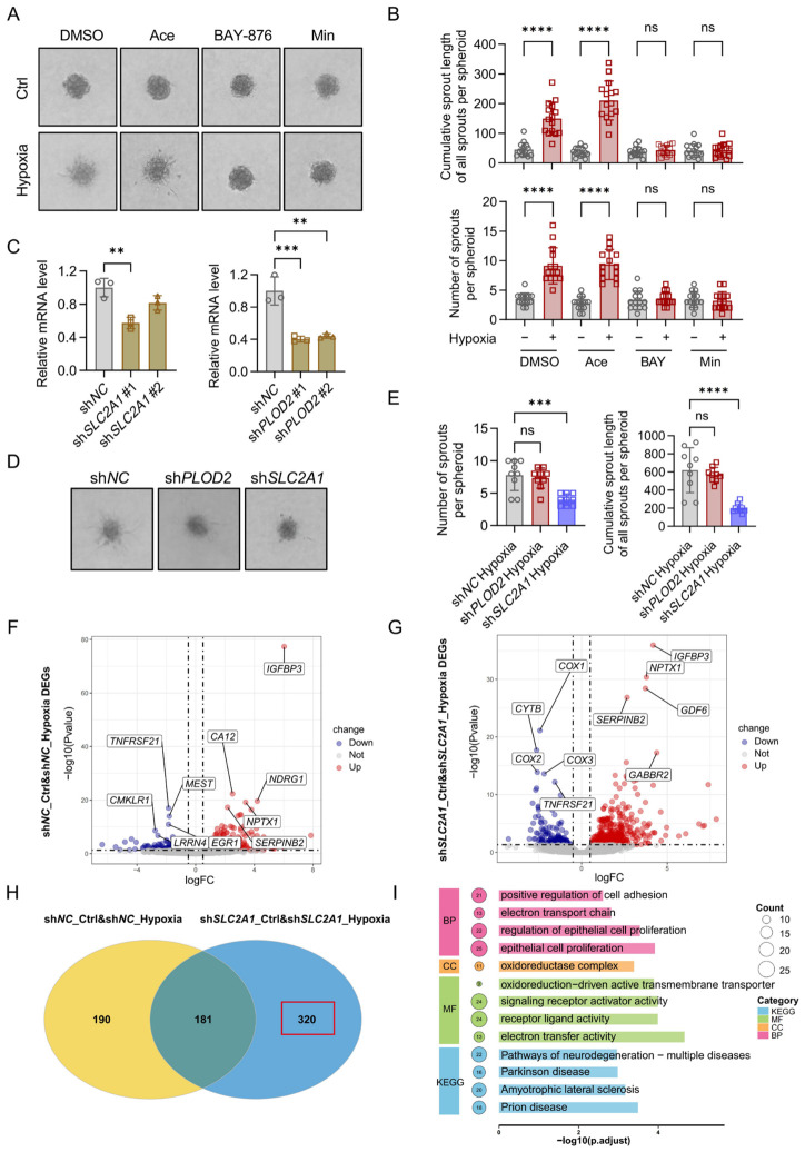
▲ SLC2A1 promotes the sprouting of endothelial cells under hypoxic conditions. (A) Representative plots showing the effects of PTGS2, SLC2A1, and PLOD2 inhibitors and DMSO on endothelial cells sprouting (Acetaminophen, inhibitor of PTGS2; BAY-876, inhibitor of SLC2A1; Minoxidil, inhibitor of PLOD2). (B) Cumulative sprout length of all sprouts and number of sprouts per spheroid in the inhibitor and control groups. (C) mRNA expression levels of shPLOD2 and shSLC2A1 in ICC-derived CAFs. (D) Representative plots showing the effects of conditioned media from shPLOD2, shSLC2A1, and control groups on endothelial cells sprouting. (E) Cumulative sprout length of all sprouts and number of sprouts per spheroid in the shPLOD2, shSLC2A1, and control groups. (F, G) Volcano plots of DEGs in control and shSLC2A1 under normal and hypoxic conditions. (H) Venn diagram showing the overlap of DEGs between the control and shSLC2A1 groups. (I) GO and KEGG functional enrichment analyses based on the uniquely expressed genes in the shSLC2A1 group. **, p < 0.01; ***, p < 0.001; ****, p < 0.0001; ns, not significant.
BAY-876 (T3713) was used as a specific SLC2A1 inhibitor administered intragastrically in an orthotopic intrahepatic cholangiocarcinoma mouse model. Using BAY-876 to inhibit SLC2A1 significantly attenuated tumor burden, demonstrated by a marked reduction in liver-to-body weight ratio and absolute tumor weight. Consistent with these reductions, in vivo bioluminescence imaging showed that total light flux in the BAY-876 group was significantly lower than in controls, indicating decreased tumor activity. Beyond effects on primary tumor metrics, SLC2A1 inhibition significantly suppressed intrahepatic metastasis, and the number of metastatic liver nodules was markedly reduced in the BAY-876 treatment group compared with the control group. The study further verified the potential efficacy of BAY-876 against tumor progression in the orthotopic intrahepatic cholangiocarcinoma model. Together, these findings characterize BAY-876 as a selective SLC2A1 inhibitor that, when administered intragastrically, reduces tumor burden, diminishes bioluminescent tumor signal, and suppresses intrahepatic metastatic spread in vivo.[3]
■ 2.2 Biomimetic nanomodulator reprograms glycolysis-driven immunosuppressive microenvironment to potentiate photothermal immunotherapy in cold tumors.
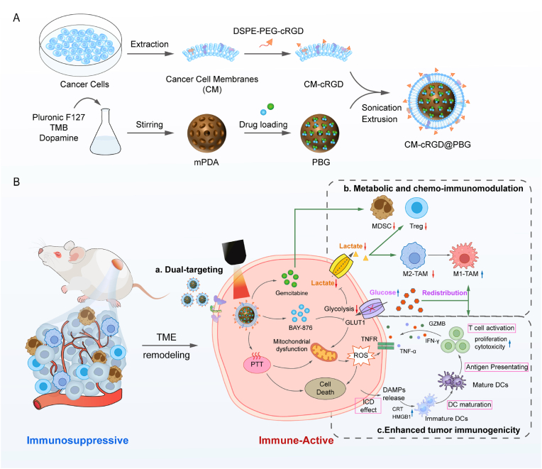
▲ Schematic illustration of the biomimetic nanomodulator-mediated synergistic “metabolic-chemo-photothermal” therapy against “cold” tumors. (A) Preparation of CM-cRGD@PBG. cRGD-modified cancer cell membranes (CM) are coated onto mPDA nanoparticles co-loaded with BAY-876 and gemcitabine (GEM). (B) Mechanism of synergistic antitumor efficacy. CM-cRGD@PBG efficiently targets and accumulates at the tumor site via homologous and active targeting. After cellular internalization, the released BAY-876 and GEM remodel the immunosuppressive tumor microenvironment (ITME) by redistributing glucose, inhibiting lactate production, and reducing immunosuppressive cell populations. Simultaneously, photothermal therapy induces apoptosis and triggers immunogenic cell death (ICD) to amplify tumor immunogenicity. This strategy reprograms the TME from an immunosuppressive to an immune-active phenotype, thereby robustly potentiating cytotoxic T lymphocyte infiltration and cytotoxicity.
BAY-876 (T3713) is described as a GLUT1 blocker and is associated with inducing metabolic stress. In cellular assays, adding TNF-α to BAY-876-treated Panc02 or LLC cells significantly promoted tumor cell death compared to TNF-α alone. In a co-culture system, flow cytometry showed that the CTL-mediated tumor cell apoptosis rate in the BAY-876 group rose to 18.6% from 9.1% in the control group, supporting that BAY-876 can significantly boost CTL-mediated killing. In tumor immune profiling, representative immunohistochemistry images revealed that BAY-876-containing groups displayed a distinct increase in M1-like macrophages and a significant decrease in M2-like macrophages. This shift from M2-like to M1-like macrophages indicated that BAY-876 effectively reduced lactate-driven M2-like tumor-associated macrophage polarization through metabolic modulation. Consistent with impaired glucose utilization, cultures in the BAY-876 group showed substantially higher glucose levels in the supernatant than controls. Together, these findings depict BAY-876 as a metabolic modulator that blocks GLUT1, elevates extracellular glucose levels consistent with reduced uptake, enhances CTL-mediated apoptosis of tumor cells, augments TNF-α-dependent cytotoxicity, and reprograms macrophage polarization toward an M1-like phenotype while diminishing the M2-like state through metabolic remodeling.[4]
■ 2.3 Regulatory role of GLUT1 in lead-induced neurotoxicity and the neuroprotective effect of Morinda officinalis oligosaccharides.
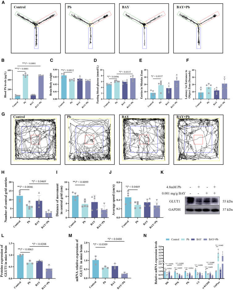
▲ Effect of BAY-876 on cognition, emotional behavior, GLUT1 expression, and glucose metabolism in Pb-exposed mice. (A) Y-maze task. (B–D) Blood Pb concentrations, brain-to-body weight ratios, and fasting blood glucose (FBG) levels (n = 5). (E, F) Number of mistake-arm entries and latency to reward-arm entry (n = 5). (G) Open field test (OFT) maps. (H–J) Number of central zone entries, central zone travel distance, and average movement speed (n = 5). (K–M) Relative protein and mRNA expression levels of GLUT1 in mouse brain (n = 3). (N) Relative mRNA expression levels of rate-limiting glucose metabolism enzymes (n = 3). The results demonstrate that Pb exposure impairs cognitive and emotional behaviors, accompanied by abnormal expression of GLUT1 and rate-limiting enzymes in glucose metabolism; administration of BAY-876 partially reverses these behavioral and metabolic abnormalities. Data are presented as mean ± SD and were analyzed using a one-way ANOVA with Tukey’s post-hoc test. Each experiment included at least three independent replicates per group. ∗p < 0.05; ∗∗p < 0.01; ∗∗∗p < 0.001. “–” indicates no specific treatment; “+” indicates treatment.
BAY-876 (T3713) was evaluated in a Pb-exposed mouse model, where it produced several measurable effects on behavior, metabolism, and neuronal structure. In systemic metabolism, fasting blood glucose levels were significantly higher with BAY-876 in Pb-exposed mice. In behavioral assays, BAY-876 decreased central area entries in the open field test, and the authors conclude that BAY-876 exacerbates anxiety-like behavior in Pb-exposed mice and further disrupts brain glucose metabolism. At the molecular level in brain tissue, BAY-876 combined with Pb further reduced GLUT1 expression compared with Pb alone. Consistent with disrupted glucose handling, the mRNA levels of key rate-limiting metabolic enzymes—hexokinase, phosphofructokinase, and α-ketoglutarate dehydrogenase—were significantly lower with BAY-876 under Pb exposure. Structural analyses of hippocampal neurons showed that BAY-876 with Pb reduced dendritic complexity, particularly within 50–80 μm of the soma, and also decreased the number of dendritic endpoints and overall spine density. Correspondingly, synaptic protein analyses revealed significantly decreased expression of drebrin (Dbn) and PSD95 at both the mRNA and protein levels, alongside reduced GABAR protein, in the BAY-876 with Pb group. Together, these findings indicate that in Pb-exposed mice, BAY-876 elevates fasting blood glucose, exacerbates anxiety-like behavior, downregulates GLUT1 and multiple metabolic enzymes, and impairs dendritic architecture with reduced synaptic-associated proteins.[5]
■ 2.4 A Biomimetic Nanoagonist for Synergistic Photodynamic/Ferroptosis/Disulfidptosis Therapy via Coinhibition of Glucose Metabolism and the SLC7A11/GSH/GPX4 Axis.
BAY-876 (T3713) functions as a GLUT1 inhibitor, and BAY-876-mediated GLUT1 inhibition disrupts NADPH generation, leading to disulfide stress, actin cytoskeletal collapse, and disulfidptosis. In SLC7A11-high head and neck squamous cell carcinoma settings under glucose starvation, ALMS1-IT1 knockdown increases the NADP+/NADPH ratio, promotes disulfide bond formation among actin cytoskeletal proteins, induces F-actin collapse, and triggers disulfidptosis; these effects are enhanced by GLUT inhibitors such as BAY-876. Consistently, in SLC7A11-high cells, BAY-876 exposure produced characteristic cysteine/disulfide metabolic changes, and sensitivity could be functionally linked to cystine dependence, SLC7A11 status, and rescue by metabolic interventions such as cystine withdrawal. In a separate context describing a protective phenotype against disulfide stress and cytoskeletal collapse, this phenotype can be further potentiated by GLUT1 inhibition with BAY-876. Together, these findings position BAY-876 as a GLUT1-targeting agent that disrupts cellular reductive capacity and promotes a cascade characterized by NADPH failure, disulfide stress, actin cytoskeletal collapse, and disulfidptosis, while also intensifying defined redox and cytoskeletal responses under glucose-limiting and SLC7A11-high conditions.[6]
■ 2.5 D-glucose uptake inhibits bovine alphaherpesvirus 1 post-binding cell process entry via inhibition of PLC-γ1 signaling in a glucose transporter 1-independent manner.
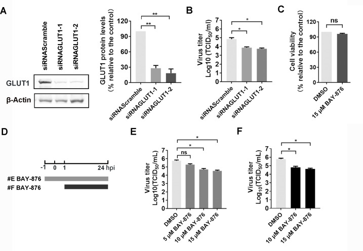
▲ (A) MDBK cells in six-well plates were transfected with either scrambled siRNA (150 pmol) or two individual siRNAs targeting GLUT1 (150 pmol each), referred to as siRNAGLUT1-1 and siRNAGLUT1-2, respectively. At 48 h post-transfection, GLUT1 protein levels were detected via Western blot, and band intensity was analyzed using ImageJ software. (B) MDBK cells in six-well plates were transfected with 150 pmol of scrambled siRNA, siRNAGLUT1-1, or siRNAGLUT1-2. After 36 h transfection, cells were infected with BoAHV-1 (MOI = 1) for 1 h. After three washes with PBS, fresh medium was replaced. At 24 hpi, virus yield in the cell cultures was measured and expressed as TCID50/mL. (C) The cytotoxicity of BAY-876 (15 μM) in MDBK cells for 24 h was analyzed by Trypan-blue exclusion test. (D) Diagram showing two different experimental conditions. (E) BAY-876 treatment from −1 to 24 hpi. (F) BAY-876 treatment from 1 to 24 hpi. (E) MDBK cells in 24-well plates pretreated with either DMSO control or BAY-876 at indicated concentrations for 1 h were infected with BoAHV-1 (MOI = 1) for 1 h in the presence of the indicated chemicals. After three washes with PBS, fresh media containing DMSO control or BAY-876 were added for further incubation. At 24 hpi, virus titers were measured and expressed as TCID50/mL. (F) MDBK cells in 24-well plates were infected with BoAHV-1 (MOI = 1) for 1 h. After three washes, fresh media containing respective chemicals were added for further incubation. At 24 hpi, virus titer was determined and expressed as TCID50/mL. The results shown are means of three independent experiments, with error bars indicating SDs. Significance was assessed with Student’s t-test (*, P < 0.05; **, P < 0.01; ns, not significant).
BAY-876 (T3713) is described as a highly potent and selective inhibitor of GLUT1 compared to other GLUTs. In cellular assays, BAY-876 significantly diminished β-catenin-dependent transcriptional activity stimulated by productive virus infection at 10 and 15 μM in MDBK cells, and it also inhibited β-catenin-dependent transcription in MDBK cells under mock infection at the same concentrations. In Neuro-2A cells, β-catenin-dependent transcriptional activity was significantly decreased by BAY-876 in a dose-dependent manner. Consistent with these transcriptional effects, the protein levels of phosphorylated β-catenin at Ser552 in MDBK cells decreased following BAY-876 exposure, irrespective of the presence of viral infection. Beyond signaling readouts, BAY-876 reduced BoAHV-1 productive growth in MDBK cells. When administered as a 1-hour pretreatment and maintained during infection, viral titers declined by approximately 1.05 and 1.22 log units at 10 and 15 μM, respectively. When applied starting at 1 hour post-infection and maintained to 24 hours, viral titers were reduced by approximately 0.95 and 1.15 log units at 10 and 15 μM, respectively. These findings collectively indicate that BAY-876 suppresses β-catenin-associated transcriptional activity and lowers phosphorylated β-catenin levels, while also decreasing BoAHV-1 viral yields in MDBK cells under multiple dosing regimens.[7]
■ 3. Conclusion
BAY-876 acts as a highly selective and potent inhibitor of the facilitative glucose transporter GLUT1, directly blocking glucose translocation across the plasma membrane. This inhibition restricts cellular glucose influx, thereby suppressing glycolysis and downstream biosynthetic and redox pathways, inducing energy and nutrient stress that modulates key metabolic sensors and growth signaling. Literature reports demonstrate that BAY-876 effectively reduces tumor burden and metastasis in intrahepatic cholangiocarcinoma models, enhances cytotoxic T lymphocyte-mediated tumor cell apoptosis, and shifts macrophage polarization from an immunosuppressive M2-like phenotype to a pro-inflammatory M1-like state. Additionally, BAY-876 exacerbates metabolic and behavioral dysfunctions in neurotoxicity models, disrupts cellular redox homeostasis to induce cytoskeletal collapse and disulfidptosis in cancer cells, and inhibits β-catenin signaling and viral replication in infected cells. Collectively, these findings highlight BAY-876’s value as a chemical tool and therapeutic candidate targeting metabolic vulnerabilities. Future research may focus on combinatorial therapies with complementary metabolic inhibitors, further exploration of immune microenvironment remodeling, and detailed mechanistic studies in neurological and infectious disease contexts.
■ 4. Q&A
Q1: What is the primary mechanism of action of BAY-876?
A1: BAY-876 selectively inhibits GLUT1 by directly blocking glucose transport across the plasma membrane, thereby reducing cellular glucose influx and suppressing glycolysis and its downstream metabolic pathways.
Q2: How does BAY-876 affect tumor progression and immune response according to the literature?
A2: BAY-876 reduces tumor burden and intrahepatic metastasis, enhances CTL-mediated tumor cell apoptosis, and shifts macrophage polarization from an immunosuppressive M2-like phenotype to a pro-inflammatory M1-like state by metabolic remodeling.
Q3: What additional biological effects of BAY-876 have been observed beyond cancer models?
A3: BAY-876 exacerbates anxiety-like behavior and disrupts brain glucose metabolism in lead-exposed mice, induces disulfide stress and cytoskeletal collapse leading to disulfidptosis in cancer cells under glucose starvation, and inhibits β-catenin-dependent transcription and viral replication in infected cells.
■ 5. Reference
[1] Siebeneicher H, Cleve A, Rehwinkel H, et al. Identification and Optimization of the First Highly Selective GLUT1 Inhibitor BAY-876. ChemMedChem. 2016;11(20):2261-2271.
[2] Macheda ML, Rogers S, Best JD. Molecular and cellular regulation of glucose transporter (GLUT) proteins in cancer. J Cell Physiol. 2005;202(3):654-662.
[3] Fan J, Tong M, Lu Y, Wang Q, Xie Y, Lin K, et al.. Single-Cell and Bulk Transcriptomics Uncover the Cellular Ecosystem of Vascular Invasion in Intrahepatic Cholangiocarcinoma.. Cells. 2026;15(11):.
[4] Zhao X, Yang Y, Niu Y, Feng J, Yuan W, Liu M. Biomimetic nanomodulator reprograms glycolysis-driven immunosuppressive microenvironment to potentiate photothermal immunotherapy in cold tumors.. Materials today. Bio. 2026;38():103195.
[5] An S, Ge Y, Liu Y, Sun Z, Chen M, Chi C, et al.. Regulatory role of GLUT1 in lead-induced neurotoxicity and the neuroprotective effect of Morinda officinalis oligosaccharides.. iScience. 2026;29(4):115342.
[6] Jiang Z, Zhao Y, Li Z, Jiang H, Lv B, Cao J. A Biomimetic Nanoagonist for Synergistic Photodynamic/Ferroptosis/Disulfidptosis Therapy via Coinhibition of Glucose Metabolism and the SLC7A11/GSH/GPX4 Axis.. ACS applied materials & interfaces. 2026;18(10):14755-14772.
[7] Li X, Ding X, Zhang N, Ma X, Fiorito F, Zhu L. D-glucose uptake inhibits bovine alphaherpesvirus 1 post-binding cell process entry via inhibition of PLC-γ1 signaling in a glucose transporter 1-independent manner.. Microbiology spectrum. 2026;14(2):e0245625.
http://www.targetmol.com
TargetMol Chemicals Inc. -
AuthorPosts

- You must be logged in to reply to this topic.